




| Class | Growth factor (PEGylated) |
| Molecular weight (Da) | variable |
| Half-life (indicative) | prolonged (days) † |
| Research status | Preclinical / RUO |
Research context (RUO): MGF (Mechano Growth Factor) and PEG-MGF are peptide variants derived from a locally produced splice variant of IGF-1. They are supplied exclusively for in-vitro laboratory research and are not intended for human or animal use.
MGF and PEG-MGF research: mechano growth factor explained
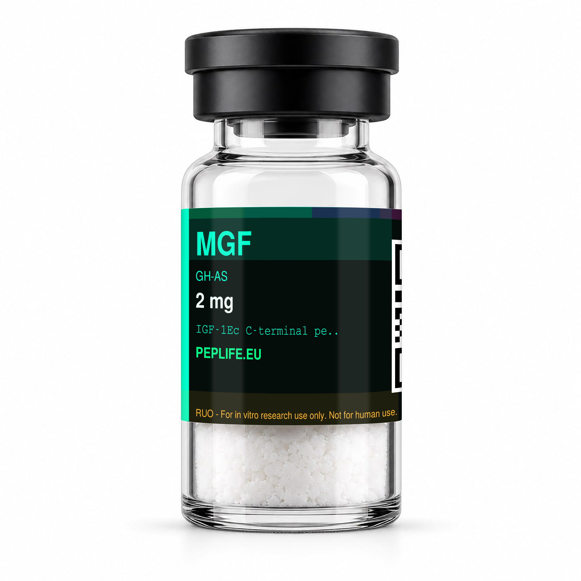
MGF and PEG-MGF research revolves around a striking discovery from muscle physiology: when muscle tissue is loaded or damaged, the cell splices the IGF-1 gene together in a different way and produces a short, locally acting variant. That variant was given the name Mechano Growth Factor, MGF for short, because its production is driven by mechanical signals. In this article we explain what MGF is, how it relates to the pegylated form PEG-MGF, and what both are studied for in laboratory studies.
What are MGF and PEG-MGF?
MGF is not a separate protein that floats around freely in the body, but a splice variant of IGF-1 (insulin-like growth factor 1). When the IGF-1 gene is read out, the cell can combine different exons; under mechanical loading the so-called IGF-1Ec isoform arises in humans (called IGF-1Eb in rodents). The distinguishing feature lies in the C-terminal part, the so-called E-domain or E-peptide. Researchers often study a synthetic peptide that corresponds to this unique C-terminal part; that is what is commonly sold as “MGF”.
The practical problem in research is that the native MGF peptide is broken down very quickly in serum and therefore has only a short window of action. For this reason, PEG-MGF was developed: the same peptide sequence to which a polyethylene glycol chain (PEG) is attached. Pegylation is an established pharmaceutical technique that increases the molecular mass, protects the peptide against enzymatic degradation and thus extends the half-life. The difference between MGF and PEG-MGF therefore lies not in the biological core, but in the stability and the duration that the molecule remains intact in a solution or model system.
How does MGF work? — the mechanism
The central hypothesis in MGF research is that the two IGF-1 splice variants fulfil different roles. According to the review work of Geoffrey Goldspink (Physiology, 2005), the IGF-1Ec/MGF variant would form the first, fast response after loading — it “kickstarts” the repair process — while the later, systemic IGF-1Ea variant is associated more with differentiation and growth. Researchers state that MGF is mainly studied in relation to the activation of satellite cells: the resting precursor cells along muscle fibres that can divide upon damage and supply new nuclei to the fibre.
Important for a fair picture: this remains a subject of scientific debate. In a minireview in Endocrinology (Matheny et al., 2010), researchers point out that there is confusion about the nomenclature and that there is “a lack of definitive evidence” for a direct, independent action of the MGF-E peptide on satellite cell activation. In other words, MGF and PEG-MGF research is active and not closed — one reason these peptides remain interesting as research material.
What are MGF and PEG-MGF studied for?
In preclinical and in-vitro studies, researchers focus on a few recurring themes:
- Satellite cells and muscle recovery: studies investigate whether the MGF-E peptide stimulates the proliferation of muscle precursor cells. In a study on human muscle progenitor cells (Ates et al., 2011), researchers reported that the MGF-E peptide activated these cells and increased their fusion potential, also in cells from older donors.
- Muscle loss in ageing and disease: researchers have associated a reduced ability to express MGF with age-related muscle loss (sarcopenia) and with muscle-wasting conditions. The expression of the splice variant is central to this.
- Mechanotransduction: how mechanical signals such as stretch, exercise and electrical stimulation drive the alternative splicing of the IGF-1 gene — the core of why MGF is called “mechano”.
- Tissue beyond muscle: the IGF-1Ec/MGF isoform has also been found in other tissues; a PLOS One study (2013), for example, described expression in the growth plate of bone.
In this context, PEG-MGF is mainly studied as a more stable model form: because it remains intact longer, researchers can observe effects over a longer time span than with the rapidly degraded native peptide.
MGF vs PEG-MGF: the difference at a glance
In short: MGF is the direct, short-acting form that mimics the fast, local repair signal as it arises in tissue right after loading. PEG-MGF is the same sequence with a PEG chain, which sharply increases the half-life and makes the molecule more suitable for experiments where a longer, more uniform presence is desired. Researchers thus choose between “fast and short” (MGF) and “slow and long-lasting” (PEG-MGF) depending on their research setup. Both fall within the broader family of the GH-axis peptides, together with related molecules such as IGF-1 LR3.
MGF and PEG-MGF in laboratory research: handling & dissolving
Both peptides are supplied as freeze-dried (lyophilised) powder and must be reconstituted before use in a research protocol. As with most peptides, bacteriostatic or sterile water is usually used for this; the precise choice and dilution depend on the experimental setup. An extensive explanation of solvents and method is in our pillar reconstitute peptides and in which liquid do you use to dissolve peptides. Reconstituted peptides are stored refrigerated; native MGF is more susceptible to degradation than the pegylated variant.
Quality & purity
Every relevant batch is independently HPLC-tested by an external laboratory; the certificate of analysis (CoA) is publicly verifiable per batch. For research material this is essential: with peptides like MGF and PEG-MGF, the purity and correct pegylation directly determine the reproducibility of experiments. We reject batches that do not meet the standard — read why in why we rejected a batch.
Frequently asked questions about MGF and PEG-MGF
What is the difference between MGF and PEG-MGF?
MGF is the native, short-acting peptide that mimics the C-terminal E-domain sequence of the IGF-1Ec splice variant. PEG-MGF is the same sequence with an attached PEG chain, which greatly extends the half-life. In research, PEG-MGF is therefore used when a longer stability is desired.
Is MGF the same as IGF-1?
Not entirely. MGF is a splice variant of the IGF-1 gene — the IGF-1Ec isoform — with a unique C-terminal E-peptide. It thus shares the same genetic origin as IGF-1, but has a different structure and is studied in research as a separate, locally acting factor.
Why is it called “mechano” growth factor?
Because the production of this splice variant in tissue is driven by mechanical signals such as stretch, loading and electrical stimulation. Researchers use the name to emphasise that it concerns a mechanically induced response.
What are MGF and PEG-MGF studied for?
Mainly for satellite cell activation, muscle repair after damage, and the role of reduced MGF expression in age-related muscle loss. The findings are attributed in scientific publications to the study, not to users.
Can I use MGF or PEG-MGF?
No. Both are supplied exclusively for in-vitro laboratory research (Research Use Only) and are not intended for human or animal use, nor diagnostic or therapeutic.
How does MGF relate to other GH-axis peptides?
MGF sits downstream in the growth hormone-IGF axis: growth-hormone-releasing peptides indirectly increase IGF-1 signalling, while MGF mimics a local splice variant of it. More context is in our pillar on growth hormone peptides.
Read more & research at Peplife
- Research MGF at Peplife
- Research PEG-MGF at Peplife
- View all GH axis peptides
- Growth-hormone peptides: the GH axis explained
- Research IGF-1 LR3 at Peplife
- Research Follistatin-344 at Peplife
Sources: Goldspink, Physiology (Bethesda), 2005 · Matheny et al., Endocrinology, 2010 (MGF minireview) · Ates et al., MGF-E peptide and human muscle progenitor cells, 2011 · IGF-1Ec/MGF in the growth plate, PLOS One, 2013
Research Use Only. All products are supplied exclusively for in vitro laboratory research. Not intended for diagnostic or therapeutic use in humans or animals, and not approved by the EMA or FDA.
HPLC-tested by an external laboratory, CoA publicly verifiable per batch and discreet EU shipping.